- Joos E, Bourgeois P, Famaey JP (June 1993). "Lymphatic disorders in rheumatoid arthritis". Seminars in Arthritis and Rheumatism. Elsevier BV. 22 (6): 392–398.
- Grada AA, Phillips TJ (December 2017). "Lymphedema: Diagnostic workup and management". Journal of the American Academy of Dermatology. 77 (6): 995–1006.
- Sleigh BC, Manna B (January 2020). "Lymphedema". StatPearls [Internet]. Treasure Island (FL): StatPearls Publishing.
- Grada AA, Phillips TJ (December 2017). "Lymphedema: Pathophysiology and clinical manifestations". Journal of the American Academy of Dermatology. 77 (6): 1009–1020.
- Martin MB, Kon ND, Kawamoto EH, Myers RT, Sterchi JM (October 1984). "Postmastectomy angiosarcoma". The American Surgeon. 50 (10): 541–545.
- Chopra S, Ors F, Bergin D (December 2007). "MRI of angiosarcoma associated with chronic lymphoedema: Stewart Treves syndrome". The British Journal of Radiology. 80 (960)
- Requena L, Sangueza OP (February 1998). "Cutaneous vascular proliferations. Part III. Malignant neoplasms, other cutaneous neoplasms with significant vascular component, and disorders erroneously considered as vascular neoplasms". Journal of the American Academy of Dermatology. 38 (2 Pt 1): 143–75, quiz 176–8.
- Publishing, Licorn (2009-10-28). "Body image and quality of life in secondary lymphedema of the upper limb". Servier - Phlebolymphology.
- "Oncology Fact Sheet" (PDF). aota.org. Retrieved 2019-09-03.
- "A Visual Guide to Lymphedema". WebMD.
- Shenoy, R. K. (2008-09-20). "Clinical and Pathological Aspects of Filarial Lymphedema and Its Management". The Korean Journal of Parasitology. 46 (3): 119–125.
- "Lymphoedema - Causes". nhs.uk. 2017-10-20.
- Jeannie Burt; Gwen White (1 January 2005). Lymphedema: A Breast Cancer Patient's Guide to Prevention and Healing. Hunter House. pp. 9. ISBN 978-0-89793-458-9.
- Choices, NHS. "IPS retired". nhs.uk.
- Brorson H, Ohlin K, Olsson G, Svensson B, Svensson H (June 2008). "Controlled compression and liposuction treatment for lower extremity lymphedema". Lymphology. 41 (2): 52–63.
Lymphedema: What is it and how does it manifest? + Treatment
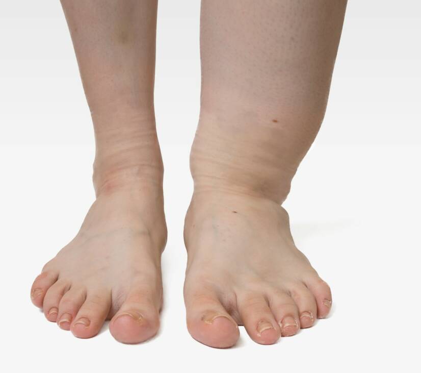
Photo source: Getty images
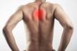
Most common symptoms
Show more symptoms ᐯ
Course
Lymphedema can occur at an early stage in secret. And so without the limb being swollen, and therefore enlarged. The first symptom may be a feeling of pressure, weakness, or fatigue of the limb. Subsequently, edema develops during a full day of exercise. However, at this point, the positioning, ie the elevated position of the limb, helps.
The third stage is characterized by persistent swelling. This is also evident in that the hole remains in the skin after being pushed with a finger. It will disappear over time. However, this condition persists for a shorter time. In the case of a greater extent of fibrotic tissue damage, the skin is stiff. And even this pressure test is not demonstrable.
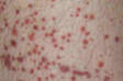
The swelling is noticeable during this period, the limb is enlarged. Paradoxically, with extensive fibrotic tissue changes, limb shrinkage occurs. The skin is stiff, movement in the joint is limited. Skin inflammation may also be associated. And even in the form of surface damage or significant ulceration. When deep ulcers develop on the surface of the skin.
The late-stage is characterized by deformities of the limb, also referred to as elephantiasis. In case of complications, worsening of lymphedema is known. In the case of primary lymphedema, swelling begins to develop in the lower limbs and progresses upwards. In the case of secondary edema, it is in the area under the disability, downwards.

Lymphedema and its treatment: lifestyle interventions and medications
Show moreLymphedema is treated by
Other names
lymphoedema