- Gardner A, Gardner E, Morley T (May 2011). "Cauda equina syndrome: a review of the current clinical and medico-legal position". European Spine Journal. 20 (5): 690–7. doi:10.1007/s00586-010-1668-3. PMC 3082683. PMID 21193933.
- "Cauda equina syndrome". Genetic and Rare Diseases Information Center (GARD). 2015. Retrieved 9 November 2017.
- "Cauda Equina Syndrome-OrthoInfo – AAOS". orthoinfo.aaos.org. March 2014. Retrieved 9 November 2017.
- Shapiro S (February 2000). "Medical realities of cauda equina syndrome secondary to lumbar disc herniation". Spine. 25 (3): 348–51, discussion 352. doi:10.1097/00007632-200002010-00015. PMID 10703108. S2CID 44975909.
- Chau AM, Xu LL, Pelzer NR, Gragnaniello C (2014). "Timing of surgical intervention in cauda equina syndrome: a systematic critical review". World Neurosurgery. 81 (3–4): 640–50. doi:10.1016/j.wneu.2013.11.007. PMID 24240024.
- Larner AJ (2006). A Dictionary of Neurological Signs (2nd ed.). [New York]: Springer Science+Business Media, Inc. ISBN 9780387262147.
- Kraemer (2009). Intervertebral disk diseases causes, diagnosis, treatment, and prophylaxis (3rd ed.). Stuttgart: Thieme. ISBN 9783131495617.
- Gardner A, Gardner E, Morley T (May 2011). "Cauda equina syndrome: a review of the current clinical and medico-legal position". European Spine Journal. 20 (5): 690–7. doi:10.1007/s00586-010-1668-3. PMC 3082683. PMID 21193933.
- Gerald L Burke, MD. "Backache from Occiput to Coccyx". Retrieved 2014-07-23.
- Eck JC (11 May 2007). "Cauda Equina Syndrome Causes". Cauda Equina Syndrome. WebMD.
What is Cauda Equina Syndrome? Causes, Symptoms and Treatment
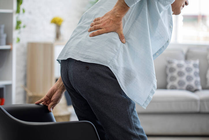
Photo source: Getty images
Cauda equina syndrome is an acute condition that occurs when the bundle of nerves below the end of the spinal cord is compressed. It causes pain and neurological damage.
Most common symptoms
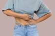
- Abdominal Pain
- Limb pain
- Nerve pain
- Lower Abdominal Pain
- Groin Pain
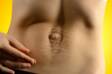
- Back Pain
- Fatigue
- Anxiety
- Urinary retention - anuria/retentiveness
Show more symptoms ᐯ
Characteristics
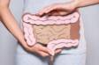
Acute compression of the tangle of nerves leaving the spinal cord is referred to as Cauda equina syndrome or Degenerative lumbosacral stenosis (DLSS).
It is an acute condition that threatens a person with serious neurological problems. Without prompt action, there is a risk that the consequences will persist.
What is Cauda eqiuna?
The name comes from Latin and it means "horse's tail".
It represents a bundle of nerves that looks like a horse's tail.
The spine is composed of vertebrae. Theyare classified by their location into segments: cervical, thoracic, lumbar, sacral, and coccygeal vertebrae.
The spine is composed of a different number of vertebrae:
Cervical vertebrae = 7 - vertebrae Cervicales - C
Thoracic vertebrae = 12 - vertebrae Thoracicae - Th
Lumbar vertebrae = 5 - vertebrae Lumbales - L
Sacral vertebrae = 5 or 6 - vertebrae Sacrales - S, they form the sacrum = Lat. os sacrum
Coccyx = 4 or 5 - vertebrae Coccygeae - Co
The vertebrae have a specific shape and function. In addition to carrying the body's weight, their role is to protect the spinal cord.
The spinal cord is located in the spinal canal, which consists of vertebral bodies and arches.
Spinal cord = Lat. medulla spinalis.
It is located from the first cervical vertebra C1. It grows more slowly during development, which is the result of reaching only the 1st to 2nd lumbar vertebrae - L1 to L2.
The spinal cord is about 40-50 centimetres long.
Cauda equina...
Further down in the spinal canal, the nerve bundle continues. It is referred to as the cauda equina. It protrudes from the end of the conus medullaris.
It contains the lumbar, and sacral nerves and coccygeal nerve. They innervate the pelvic organs, lower limbs, namely the motor (movement) component of the hips, knees, ankles, legs, but also anal sphincters, and sensory and parasympathetic innervation, for example for the bladder.
Pathological compression (compression of the cauda equina) results in a condition called the cauda equina syndrome.
Cauda equina is formed by nerve roots:
L1 to L5 + S1 to S5.
The roots L4 to S4 together form the plexus sacralis - sacral plexus.
The sacral plexus is the largest tangle of the human body.
The sciatic nerve comes out f it - the largest nerve of the sacral bundle.
Nervus ischiadicus runs further down to the lower limb.
Sciatica - sciatic nerve inflammation is a well-known associated condition.
The intervertebral disc plays a major role in the cauda syndrome
Intervertebral discs = Lat. disci intervertebrales. The name comes from Latin.
These are flexible shock absorbers that fill the space between the vertebrae. They are adapted to the shape of the vertebrae, copying and lining them so as not to reach outside this area.
The discs have several functions:
- damping/absorbing shocks and vibrations during the body's movement
- stabilise the spine
- maintain balance
- they balance tension and pressure
- they participate in the movement of the spine - bending and turning
The physical action and the load are distributed evenly over the entire area of the vertebrae.
Depending on the location, they have adapted to their function as well as their shape and size. The narrowest are in the cervical spine and the largest in the trunk area.
Intervertebral discs are not found between all vertebrae.
They are in spaces C2 - C3 to L5 and S1.
There are 23 discs.
Its total length is 20-25% of the total length of the spine.
From the structural point of view, they consist of three important structures, namely:
- fibrous ring - anulus fibrosus
- is the outer part of the disc
- is made of several laminae, roughly 15 - 20 of them
- the laminae are arranged in circles, like an onion
- between the laminae there is water and elastin fibers
- nucleus - nucleus pulposus
- the central pasrt of the disc
- it has a gel-like consistency - 40 %
- is harder than the fibrous ring
- contains water, protein and collagen fibres
- water content after birth 90%
- decreases with age and after the age of 50 it is 70%
- does not contain blood vessels or nerves
- vertebral endplate
- it is hyaline cartilage about 1 mm thick
- forms the boundary between the disc and the body of the vertebra
- participates in the exchange of nutrients and waste in the plate
The disc contains a blood supply only to a certain part of the ring. The centre has no vessels and its nutrition and drainage of waste substances occurs through the environment.
The flow of water is ensured mainly during movement.
In this case, the intervertebral discs are compressed and released. This alternating load expels the fluid - creep phenomenon, helps to absorb and expel fluid.
Imagine fluid being absorbed by a sponge.
Lack of exercise and a sedentary lifestyle are behind the poor nutrition of discs, which contributes to pathological processes.
The discs lose water over time, their height decreases, which has a negative effect on the stability of the entire segment. They do not distribute physical forces evenly. The ring is disrupted, which can lead to disc herniation.
Degenerative diseases of discs and vertebrae:
Osteochondrosis
Spondylosis
Spondyloarthrosis
Caude equina syndrome is..
It is a less common disease, but, more seriously, mainly from a neurological point of view.
Compression of the tangle protruding from the lumbar spinal cord disrupts neurological functions. The motor, ie movement and sensory components are affected.
Caude equina syndrome - Lat. syndroma caudae equinae - SCE.
The condition affects the folliwng areas:
- genitals
- anus
- inside area of things
- calves
- legs
Neurological disorders of the "saddle anesthesia" type.
Neurological deficit, ie insufficient function, can develop bilaterally, but also on the one hand. The course and development of difficulties is individual and may not occur in everyone in the same form.
When evaluating the syndrome according to its origin and course, the affected patients are divided into three groups, namely:
1. with a sudden onset of symptoms leading to cauda equina syndrome,
2. patients have already experienced back pain and radicular syndrome in the past,
3. with bilateral radiculopathy and with the final development of cauda syndrome.
In most cases, the syndrome is the result of oppression created by extensive disc herniation. In this case, surgery is indicated, the aim of which is to remove the oppression of the nerve mesh.
Causes
There can be several causes of kaudy equina syndrome. It most often results from damage to the intervertebral disc.
=
In other words, the displacement of the intervertebral disc due to herniation or separation of a part of the disc in the area of the lumbar spine below the level of the vertebra L2.
Disc herniation is a state of disruption of the outer layer of the intervertebral disc, or the ring. The laminae of the ring burst and the inner core of the disk penetrates through the resulting crack.
This phenomenon is also referred to as disc extrusion.
The disc extends beyond the vertebra, it is the source of spinal cord or spinal nerve compression or its root (radix). Radicular irritation occurs - radiculopathy.
Or, disc extrusion with sequestration. This involves the complete separation and disintegration of a piece of disk out of the plate.
Learn more about damaged discs:
Intervertebral disc dislocation.
However, not every disc herniation, even on a larger scale, in the area of the lumbar spine causes cauda syndrome.
Information is provided that the exact cause is therefore not fully clarified. Damage to spinal nerve cells has a role to play.
In addition to a damaged plate, the oppression can also create, for example:
- spinal canal stenosis - spinal stenosis
- vertebral instability - spondylolisthesis
- abscess in the lumbar spine
- vertebral and disc inflammation - spondylodiscitis
- injury and fracture
- postoperative condition, after spinal surgery
- epidural hematoma - blood flow in the area under the cauda membrane
- rheumatic disease
- tumor and spinal metastasis
- circulator disorder, insufficient blood supply
Symptoms
The symptoms are based on a disorder of nerve function, which results from pressure or compression.
Neurological problems are observed in the above mentioned saddle anesthesia, and therefore in the area of the genitals, rectum, thighs, calves and legs.
There is pain, its radiating pain, paresthesias, which are unpleasant sensations in the sense of tingling and other impaired sensitivity or weakness and weakening muscles. Impaired bladder or rectal function, as well as sexual function, may be present.
The main symptoms include:
- severe back pain, burning
- vyžarovanie bolesti do zadku a dolnej končatiny
- sensitivity disorder in the given dermatome, ie in the area of innervation by the corresponding nerves
- hypoaesthesia - decreased perception of skin sensitivity
- anesthesia - complete numbness of the skin, loss of sensation
- motor disorder, ie momentum, paresis (weakening) to plegia (complete paralysis)
- reflex disorder
- urinary disorders and stool control - urinary and stool incontinence
- sexual dysfunction
- in a more severe condition, paraparesis, i.e. paralysis of the lower limbs
A similar issue affects two other diseases:
1. epiconus syndrome, wherein the epiconus is the area just above the end of the spinal cord, approximately at the height of the L1 vertebra. This is an impairment of the spinal segment L4 - S2.
2. conus syndrome, the end part of the spinal cord, after which the cauda equina continues. The conus is located at the L1 site and the intervertebral disc area between the L1 and L2 vertebrae. This is the area of the spinal segment S3 - S5.
The spinal cord cone has a blood supply from the epiconus, so the epiconus syndrome occurs along with the cone syndrome.
Table: Symptoms of both the epiconus and the conus of the spinal cord
| Epiconus syndrome | Conus syndrome |
weak muscle paralysis
| mainly: sphincter disorder - stool incontinence, urinary retention |
sensitivity disorder in the area
|
sexual dysfunction:
|
| partial sphincter control failure | limb movement is maintained |
| sexual dysfunction |
|
| impaired sweat gland function, resulting in easier pressure sores |
sensory dysfunction, saddle hypoaesthesia
|
Diagnostics
Diagnosis is based on the patient's medical history and clinical picture, when a person describes their problems, the time of onset, what preceded them and, in general, all the associated difficulties.
Neurological examination is important. This includes examination of reflexes, ability to maneuver, limb motility and skin sensitivity.
Imaging methods are needed to reveal the cause, especially:
- CT
- MRI
- PMG
- EMG
- X-ray
It is important to identify the cause of the cauda equina syndrome and distinguish it from other causes. This is called a differential diagnosis. The cause may be a neurological disease.
Course
There can be different courses of the disease.
Although cauda equina syndrome is described as an acute disease (or subacute - mild, gradual, less sudden), it can be preceded by chronic back pain in the lumbar or sacral region - lumbalgia or lumbischalgia.
The symptoms are individual and the overall course depends on several factors.
It generally describes the onset of a sensory disorder in the genitals, around the rectum. At the same time, the radiation moves through the area of the sciatic muscles, ie the buttocks, to the inner thighs up to the calves and legs.
In addition to back pain, paresthesias, and therefore tingling or tingling (or other unnatural sensations), occur. The pain also spreads to the site of innervation through the damaged nerves.
There is also muscle weakness, weakness in the lower limbs. Dysfunctions of the sphincter, faecal incontinence or urinary incontinence may also be associated.
Similarly, erectile dysfunction.
The symptoms tend to be asymmetrical, i.e. one-sided, and it is not a given that they must develop to the same extent and intensity in each affected person.
Question:
What is the prognosis in patients with cauda equina syndrome?
In the case of this disease, various pieces of information are given.
In some cases, the disorders of sensitivity or muscle strength disappeared within a few hours or over a longer period of time. However, there are also cases of lasting consequences.
These can be in the form of persistent pain or recurrence of pain attacks, or varying degrees (mild, moderate or severe) impaired sensitivity or muscle weakness to motor deficit. The same is true for the treatment of sphincter and sexual disorders.
How it is treated: Cauda Equina Syndrome
Treating cauda equina syndrome: Is movement, exercise or surgery enough?
Show moreCauda Equina Syndrome is treated by
Other names
syndroma caudae equinae, Degenerative lumbosacral stenosis (DLSS)
Interesting resources

Bc. Lukáš Tóth
Healthcare worker
The secondary medical school in Nitra gave me the basis for my career in the field of health and diseases. Thanks to it, I worked for 2 years in the traumatology clinic and outpatient clinic at the Nitra Hospital. Since 2006 I was employed in the emergency medical service, where I stayed until 2017.
I completed my bachelor's degree at the University of Constantine the Philosopher in Nitra in the field of emergency health care. The bachelor's degree allowed me to continue my mission as a paramedic. In the meantime, I got a job at the emergency line 155. I have been working in pre-hospital health care until today.
I had an interest in people, health and even diseases in my childhood, which gave me the prerequisite to pursue this topic in adulthood. Studying and acquiring new information in practice provided me with a great basis for writing professional texts, in the form of articles that can be understood by ordinary people. Thus, my interest in the Health Portal has a solid foundation in years of practice and personal interest. Similarly, I am also interested in healthy eating, nutrition and overall healthy lifestyle. I fill my free time with family and sports.
View all articles by the same author